Let the structural symphony begin
Like other structural biologists, Eva Nogales works in extraordinary times. The University of California, Berkeley, faculty member now has the tools to tackle important questions about cells’ molecular machinery that would have been impossible to answer just a few years ago.
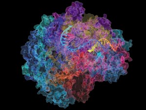
A recent project with Berkeley colleague Jennifer Doudna, the molecular biologist who co-pioneered the CRISPR–Cas9 gene-editing method, is a case in point. Both were intensely interested in the R-loop, a structure made of nucleic acids that forms in cells in many situations, but also just before DNA is snipped by CRISPR–Cas9. Nogales and her team revealed an R-loop in Streptococcus pyogenes bacteria, and from the near-atomic-resolution images, deduced how the Cas9 enzyme opens up the DNA conformation at specific sites and makes them accessible to CRISPR’s molecular scissors1.
The work is remarkable for the speed with which the scientists assigned a function to the structure, but also because they arrived at the solution by combining imaging methods — an increasingly popular approach in structural biology. For more than a century, the field’s premiere method has been X-ray crystallography. But some biomolecules are simply too big or small to crystallize, and the technique doesn’t work on others. And some biomolecules change shape or orientation as they work, which isn’t captured by static crystallization.
Read more in my first tech feature for Nature.